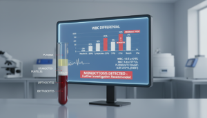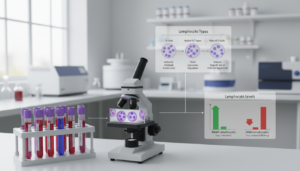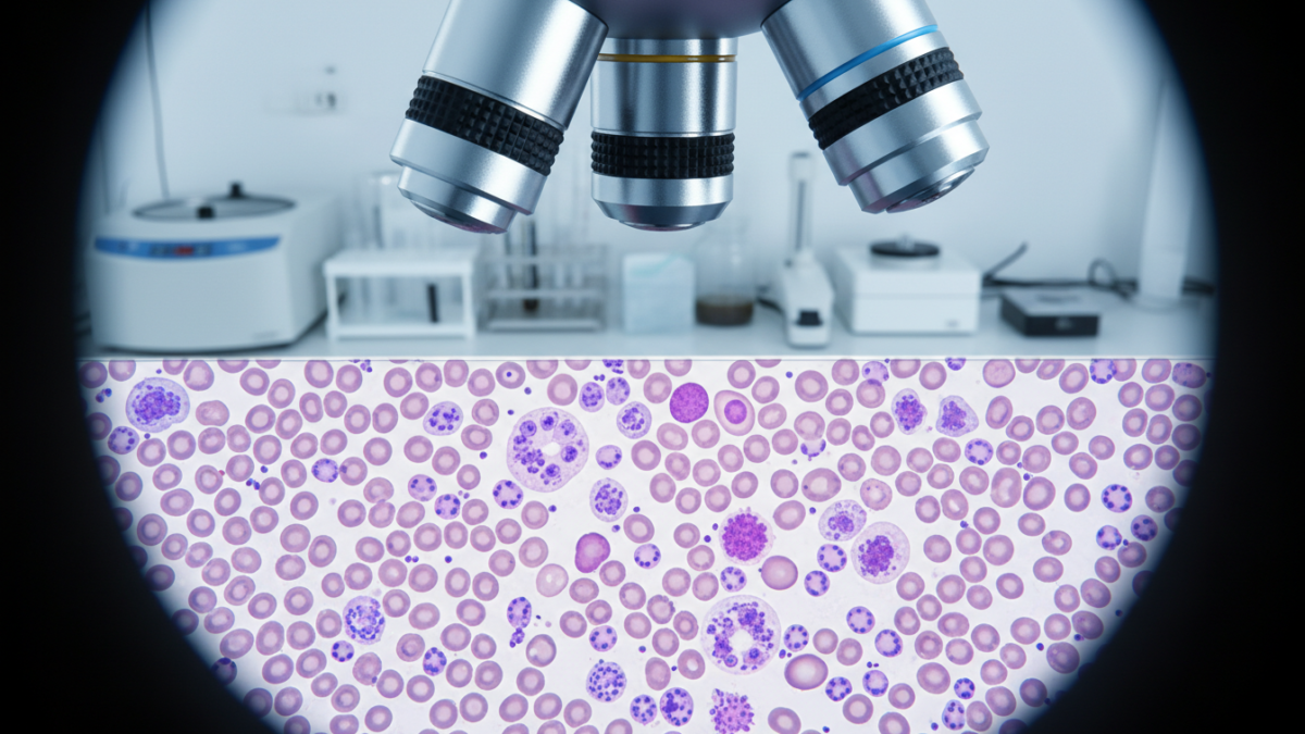
A single drop of blood has about 5 million red blood cells, 7,000 white blood cells, and 250,000 platelets. These can be seen under a microscope in a peripheral blood smear. This test has been used for over a century in hematology labs in the U.S.
A peripheral blood smear is made by spreading blood on a glass slide and staining it. Then, it’s looked at closely under a microscope. It shows the size, shape, color, and how cells are spread out. It’s also known as a blood film examination or a blood cell morphology test.
The test looks at three main types of cells. Red blood cells carry oxygen, white blood cells fight infections, and platelets help blood clot. Looking at these cells under a microscope can find problems that machines might miss.
But, a peripheral blood smear can’t diagnose a condition by itself. Doctors use it along with other tests and patient history to understand what’s going on.
Key Takeaways
- A peripheral blood smear examines red blood cells, white blood cells, and platelets on a stained glass slide under a microscope.
- The test is known by several names, including peripheral blood film, manual differential, and blood cell morphology.
- Blood film examination can detect cell abnormalities in size, shape, and color that automated analyzers may not identify.
- Microscopic blood analysis serves as a complementary tool and does not provide a standalone diagnosis.
- Wright’s stain is the standard staining method used in most clinical laboratories in the United States.
- Results from a blood smear are interpreted in combination with other laboratory tests and clinical findings.
Understanding Peripheral Blood Smears
A peripheral blood smear is a key tool in checking blood cells. It lets doctors see blood cells up close. This helps them understand what’s happening in the blood at the moment.
Definition of Peripheral Blood Smear
A blood smear is a lab test where blood is spread on a glass slide. The slide is stained to show cells under a microscope. Each slide has millions of red blood cells, thousands of white blood cells, and hundreds of thousands of platelets.
Doctors look at these cells for size, shape, color, and how they’re spread out. They might look at them by hand or with a computer system.
Purpose of a Blood Smear Test
The main goal of a blood smear test is to find problems that other tests might miss. It helps in several ways:
- Checking red blood cells for issues like anemia or sickle cell disease
- Looking at white blood cells to see if there are any odd ones out
- Checking if there are enough platelets and if they’re working right
“The blood film remains indispensable for morphologic assessment and should be reviewed whenever automated results are flagged.” — American Society of Hematology, 2023
This test gives doctors important info to make decisions. It shows a snapshot of the blood, helping to guide further tests.
The Process of Performing a Blood Smear
Creating a peripheral blood smear has two main steps. First, we get a good sample through a blood collection method. Then, we prepare the slide and stain it. Each step is very important to get accurate results.
Sample Collection Techniques
The most common way to get blood is through venipuncture. A phlebotomist uses a small needle to take blood from your arm. The blood goes into a tube with a special liquid to stop it from clotting.
For some patients, a finger prick is used. Newborns get their blood from a heel prick. The Clinical and Laboratory Standards Institute (CLSI) gives rules for each method.
Smear Preparation and Staining
First, a small amount of blood is put on a slide. Then, a spreader slide is moved over it at an angle. This makes a thin film on the slide.
After drying, we use Wright’s stain. This stain makes the cells stand out. It helps us see the details of the cells under a microscope.
| Slide Zone | Cell Appearance | Suitability for Examination |
|---|---|---|
| Thick area (near drop site) | Overlapping cells, distorted morphology | Not suitable |
| Optimal monolayer zone | Cells appear singly with central pallor | Ideal for evaluation |
| Feathered edge | Flattened cells lacking central pallor | Not suitable |
Finding the right spot to look at is key. We look at the area where red blood cells are alone and look normal.
Components of a Blood Smear
A peripheral blood smear shows three main blood cell types. Each has its own shape. Looking at these shapes under a microscope helps doctors diagnose diseases.
Red Blood Cells (Erythrocytes)
Erythrocytes are the most common cells seen. They look like diskocytes and are about 7.5 micrometers wide. They are smaller than small lymphocytes.
Hemoglobin is found around the edges of the cell. This creates a pale center. Changes in these cells can mean there’s a problem.
White Blood Cells (Leukocytes)
Leukocytes are the immune cells seen on a smear. There are five types made in the bone marrow:
- Neutrophils
- Lymphocytes
- Monocytes
- Eosinophils
- Basophils
Each type has its own shape and color. This helps doctors tell them apart under a microscope.
Platelets (Thrombocytes)
Thrombocytes are tiny, measuring 1–3 micrometers. They help stop bleeding. If they are bigger than 3 micrometers, they are called megathrombocytes.
Usually, less than 5% of thrombocytes are big. This is normal.
| Blood Cell Types | Approximate Size | Primary Function | Key Morphologic Feature |
|---|---|---|---|
| Erythrocytes | 7.5 μm | Oxygen transport | Biconcave disk with central pallor |
| Leukocytes | 7–20 μm (varies by subtype) | Immune defense | Visible nucleus with variable granules |
| Thrombocytes | 1–3 μm | Hemostasis and clotting | Small granular fragments without nucleus |
Common Findings in Blood Smears
Peripheral blood smear results show many different cell types. Some look healthy, while others suggest disease. It’s important to know the difference between normal and abnormal cells.

Normal Blood Composition
Healthy red blood cells are shaped like a disk and are about 7.5 μm wide. They have a light area in the middle that takes up 30–45% of the cell. These cells are called normocytic and normochromic.
Platelets are small, between 1–3 μm. Neutrophils have nuclei with two to five lobes.
Abnormalities Indicating Disease
Changes in red blood cell size and color can hint at disease. Anisocytosis means the cells are not all the same size. Poikilocytosis means they are not all the same shape.
There are many specific shapes that mean different things:
- Spherocytes — round, dense cells without a light area in the middle
- Target cells (codocytes) — have a light ring around a dark center
- Sickle cells (drepanocytes) — look like crescents
- Schistocytes — broken red blood cells
- Bite cells — have a hole on one side
Inclusion bodies like Howell–Jolly bodies, basophilic stippling, and Pappenheimer bodies can point to certain diseases. Rouleaux formation, where cells stack, often means there’s too much protein in the blood.
| Abnormality | Description | Clinical Association |
|---|---|---|
| Macrocytes | Diameter >9 μm | Megaloblastic anemia, B12/folate deficiency |
| Microcytes | Diameter <6 μm | Iron deficiency, thalassemia |
| Hypochromic cells | Central pallor >50% | Iron deficiency anemia |
| Acanthocytes | 3–7 irregular projections | Abetalipoproteinemia, severe liver disease |
| Echinocytes | 10–15 evenly spaced projections | Uremia, pyruvate kinase deficiency |
When is a Peripheral Blood Smear Ordered?
A peripheral blood smear is not ordered randomly. Certain signs guide doctors to order this test. It usually comes after an abnormal complete blood count (CBC) shows odd cell numbers or sizes.
The smear lets doctors see cell shape, color, and structure under a microscope.
Symptoms Leading to Tests
Certain symptoms make doctors order a blood smear. These signs might show a problem with blood or infections:
- Persistent fatigue or unexplained weakness
- Jaundice (yellowing of the skin or eyes)
- Frequent nosebleeds or unusual bleeding episodes
- Easy bruising without apparent cause
- Unexplained fever or bone pain
- Swollen lymph nodes
Travel to areas with parasites is another reason for a smear. People with fever, chills, body aches, nausea, or fatigue after traveling might need a test. This is to find organisms like Plasmodium (malaria) or Babesia (babesiosis).
Routine Screening vs. Specific Diagnosis
It’s key to know the difference between routine checks and specific tests.
| Purpose | Clinical Context | Frequency |
|---|---|---|
| Routine Monitoring | Patients under treatment for anemia or blood cancers | At scheduled intervals during therapy |
| Specific Diagnosis | New clinical symptoms suggesting hematologic disease | As indicated by abnormal CBC or acute presentation |
| Parasitic Screening | Travel history to malaria- or babesiosis-endemic areas | Upon symptom onset post-travel |
Spotting symptoms early and knowing when to order a blood smear is vital. It helps in quick and accurate diagnosis. Blood smear tests are key in checking blood health.
Understanding Blood Smear Results
After a blood smear is looked at under a microscope, a detailed report is made. This report talks about the look of red and white blood cells, and platelets. It’s important to compare these cells to known standards to understand the results.
These results are more meaningful when we think about the patient’s history and symptoms. We also look at other lab tests to get a full picture.
Interpreting Cell Counts
A manual white blood cell count tells us about five main types of cells. It shows how many of each type there are. By looking at these numbers, we can understand how they compare to each other.
Platelet counts are guessed by looking at certain areas under the microscope. Seeing 8–15 platelets in each area means the count is likely normal.
| WBC Type | Normal Adult Range (%) | Primary Function |
|---|---|---|
| Neutrophils | 40–70% | Bacterial defense |
| Lymphocytes | 20–40% | Immune regulation |
| Monocytes | 2–8% | Phagocytosis |
| Eosinophils | 1–4% | Parasitic and allergic response |
| Basophils | 0.5–1% | Inflammatory mediator release |
Identifying Cell Abnormalities
Red blood cells are checked for size, shape, and color. In healthy people, most red cells look normal. But, if more than 2% look different, it might mean something is wrong.
Looking at red blood cells helps us find problems. For example, seeing target cells or schistocytes can tell us about certain health issues. But, one smear alone can’t tell us everything. We need to look at other lab results and the patient’s symptoms to understand what’s going on.
Conditions Detected by Blood Smears
A peripheral blood smear is key for spotting many blood disorders. It shows cell shape, size, and how they’re spread out. This helps doctors find diseases, from mild to severe.
Anemia
Anemia is common in blood tests. Each type shows different signs:
- Iron deficiency anemia — small, pale red blood cells
- Megaloblastic anemia — big, oval red blood cells
- Hemolytic anemia — broken red blood cells
- Sickle cell disease — curved red blood cells
- Thalassemia — small red blood cells with special shapes
Leukemia
Too many or too few young white blood cells can mean leukemia. This calls for a bone marrow test right away.
Infections
Infections show up on blood smears. Bacterial infections make white blood cells look different. Viral infections cause special lymphocytes. Parasites can hide inside red blood cells.
| Condition | Key Smear Findings | Associated Cell Type |
|---|---|---|
| Iron Deficiency Anemia | Hypochromic microcytes | Red blood cells |
| Acute Leukemia | Blast cells, immature forms | White blood cells |
| Bacterial Sepsis | Toxic granulation, Döhle bodies | Neutrophils |
| Malaria | Intraerythrocytic parasites | Red blood cells |
| Sickle Cell Disease | Sickle cells, poikilocytes | Red blood cells |
Spotting blood disorders through smears helps doctors plan next steps. This includes more tests and seeing specialists.
Comparison with Other Blood Tests
A peripheral blood smear is a test used in blood studies. It helps doctors understand blood health. Each test has its own purpose, and knowing how they work helps doctors make the right choice.
Full Blood Count (FBC)
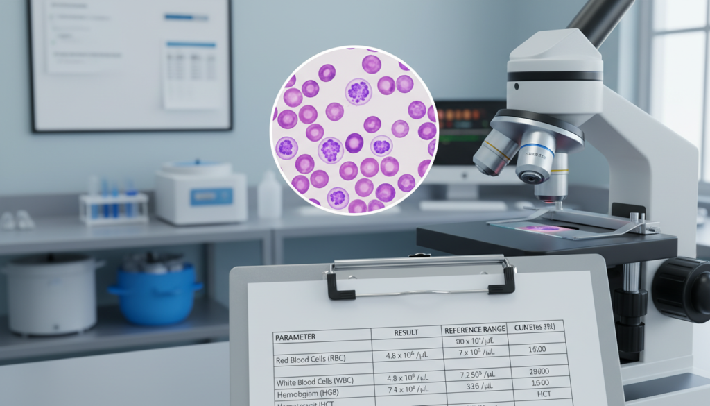
The complete blood count (CBC) is a test that counts blood cells. It’s done during checkups. It gives numbers for red and white cells and platelets.
When comparing CBC to blood smear, the main difference is what each shows. CBC finds abnormal counts. But, a blood smear shows the shape and size of cells, which machines can’t see. A study found that a blood smear was ordered in 47.4% of CBC cases. It helped find more problems (p=0.012).
- CBC was ordered in 82.7% of initial cases (373/451)
- PBS review led to bone marrow biopsy in 27.8% of further tests
- RBC disorders made up 51% of referrals, with PBS more often used (p≤0.001)
Bone Marrow Biopsy
A bone marrow test takes tissue samples. It’s used when diseases like leukemia are suspected. It shows the marrow’s structure, like cell shapes.
The blood smear is less invasive and is often the first test. In a study, all 10 bone marrow biopsies after smear review found a diagnosis. But, only 43.8% (7/16) of biopsies without smear review did.
| Feature | CBC | Blood Smear | Bone Marrow Biopsy |
|---|---|---|---|
| Data Type | Quantitative (cell counts) | Qualitative (cell morphology) | Histologic (tissue architecture) |
| Invasiveness | Minimal (venipuncture) | Minimal (venipuncture) | Moderate to high (needle biopsy) |
| Diagnostic Yield After PBS Review | N/A | Guides further testing | 100% specific diagnosis |
| Diagnostic Yield Without PBS Review | N/A | N/A | 43.8% specific diagnosis |
| Primary Clinical Role | Initial screening | Confirmatory morphologic assessment | Definitive marrow evaluation |
Preparing for a Blood Smear Test
Getting ready for a blood smear test is easy. You don’t need to follow any special diet or fast for the test alone. But, if you’re getting other tests too, you might need to fast for a few hours. Always check the instructions from your doctor before your visit.
Pre-test Instructions
There’s not much to do before a blood smear test. Just remember a few things:
- No fasting needed for the test alone
- Fasting might be required with other tests
- Wear a short-sleeved shirt for easy access
- Tell the phlebotomist about any bleeding issues or meds
What to Expect During the Test
The blood smear test is quick, taking less than five minutes. First, a band is put on your arm to make the veins stand out. Then, the phlebotomist cleans the area and draws blood with a needle.
| Stage | Description | Duration |
|---|---|---|
| Tourniquet application | Elastic band placed on upper arm | ~30 seconds |
| Site cleaning | Antiseptic applied to puncture area | ~15 seconds |
| Blood draw | Needle insertion and sample collection | 1–3 minutes |
| Post-draw care | Bandage applied with light pressure | ~30 seconds |
There’s also a finger prick method for blood collection. You might feel a bit of pain when the needle goes in. But, the site might be a bit sore or bruised for a short time. You can go back to normal activities right after the test.
Risks and Considerations
Every test has some risks for patients and doctors. Knowing about blood test risks and lab details helps set clear expectations. This includes how reliable the results will be.
Potential Risks of Blood Sampling
Getting a peripheral blood smear is considered low-risk. The main risks are a bit of pain where the needle goes in and small bruises. These usually go away in a few days without needing treatment.
“Venipuncture remains one of the safest and most routinely performed procedures in clinical medicine, with serious complications occurring in fewer than 1% of cases.” — American Society for Clinical Pathology
But, there are rare risks like infection, swelling, or feeling dizzy for a short time. People with bleeding problems might see more bleeding at the site.
Accuracy of Results
How accurate a smear is depends on many things during making and staining. Some lab steps can cause artifactual changes. These might look like real problems:
| Artifact Type | Cause | Appearance on Smear |
|---|---|---|
| Echinocytes | Hypertonic or alkaline staining solutions | Crenated red blood cells with uniform spicules |
| Stomatocytes | Acidic staining conditions | Red cells with mouth-shaped central pallor |
| Cytoplasmic vacuoles | EDTA anticoagulant exposure | Clear vacuoles in granulocyte cytoplasm |
| Basket cells | Fragile lymphocytes ruptured during spreading | Smudged nuclear remnants lacking intact cytoplasm |
| Platelet clumping | EDTA-induced aggregation | Falsely decreased platelet count estimation |
Neutrophils and monocytes often gather at the slide’s edge. This can mess up the test if not done right. It’s key to do both making and checking the smear correctly to get accurate results.
Finding a Specialist for Diagnosis
When a blood smear shows odd results, seeing a blood disorder expert is key. These doctors know how to read complex smear results. They also look at blood counts and red cell data. Getting a correct diagnosis often needs their skill.
Hematologists and Their Role
A hematologist checks blood smear details and lab data closely. Walker, Hall, and Hurst’s Clinical Methods say this is vital for diagnosing blood disorders. A specialist can tell apart conditions that look similar but need different treatments.
During a hematologist visit, they do several important things:
- They expertly look at red, white, and platelet cells.
- They match blood smear results with what the patient is feeling.
- They decide if more tests, like a bone marrow biopsy, are needed.
Importance of Follow-up Consultations
For patients with long-term blood disorders, regular check-ups are key. Blood smears are checked often to see how the disease is doing. This helps catch problems early.
| Condition | Follow-up Care Focus | Typical Monitoring Frequency |
|---|---|---|
| Polycythemia Vera | Red cell mass and viscosity changes | Every 3–6 months |
| Multiple Myeloma | Rouleaux formation and plasma cell presence | Every 1–3 months |
| Aplastic Anemia | Pancytopenia severity and response assessment | Every 2–4 weeks initially |
| Hairy Cell Leukemia | Abnormal lymphocyte detection | Every 3–6 months |
After visits, patients usually get their test results by email or mail. Each visit lets doctors check smear results again. They do this with new information about the patient’s health.
Advances in Blood Smear Technology
The field of hematology has changed a lot in recent years. Now, we have faster and more standard ways to check blood smears. These changes help at every step, from getting the sample to classifying cells and reporting results.
Automation and Digital Imaging
Now, machines can quickly check lots of blood samples. They find odd cells for experts to look at. This makes things faster and more consistent everywhere.
Digital images of cells are a big step forward. High-tech cameras on microscopes take clear pictures of blood cells. These pictures can be shared, used in teaching, and help in research. A study by Barbara Bain in the New England Journal of Medicine shows how these systems improve diagnosis.
| Feature | Manual Microscopy | Automated Digital Systems |
|---|---|---|
| Processing Speed | 15–30 minutes per slide | 3–5 minutes per slide |
| Standardization | Operator-dependent | Algorithm-driven consistency |
| Remote Consultation | Not supported | Supported via digital image sharing |
| Detection of Subtle Abnormalities | Highly effective with experienced staff | Requires manual confirmation |
Future Trends in Hematology Testing
New things are coming in lab tech, like:
- Artificial intelligence for cell recognition
- Automated counts with digital images
- Easy connection with electronic health records
Even with these new tools, manual checking of blood smears is key. Finding small cell problems needs skilled people.
Patient Experiences and Testimonials
Peripheral blood smear testing has helped many people. It has changed how doctors diagnose diseases. This has led to better care and outcomes for patients.
Real Stories from Patients
Case reports show how blood smears help doctors make decisions. For example, people with hereditary spherocytosis are found through a blood smear. This helps them get the right treatment early.
People who travel and get sick can be quickly tested for diseases like malaria. Those with myelofibrosis get help sooner because of blood smear results. These stories highlight the importance of blood smears in diagnosis.
| Condition | Key Smear Finding | Clinical Impact |
|---|---|---|
| Leukemia | Immature blast cells | Prompt oncology referral |
| Sickle Cell Disease | Irreversibly sickled cells | Early preventive care initiation |
| Vitamin B12 Deficiency | Hypersegmented neutrophils | Prevention of neurologic complications |
| Malaria | Intraerythrocytic parasites | Rapid species identification |
The Importance of Early Diagnosis
The Cleveland Clinic says early diagnosis is key. For example, leukemia can be caught before symptoms show. This means patients can get treatment sooner.
Early detection through blood smears is vital. It helps doctors spot problems quickly. This is a big part of good hematologic care.
Frequently Asked Questions about Blood Smears
A blood smear FAQ section answers common questions about this test. It helps patients and healthcare professionals understand the process better. This can make them feel more at ease about the test.
Common Concerns About the Procedure
Getting a blood smear is not very painful. It only hurts for a few seconds when the needle goes in. The test must be done by a professional in a clinic, not at home.
Results come back quickly, in a few hours or days. In the US, most insurance covers the test. This includes the cost of getting the blood, testing it, and any doctor visits.
Clarifying Misconceptions about Results
Many think a blood smear can diagnose a disease by itself. But, it’s not that simple. Doctors look at the smear, a complete blood count, symptoms, and more tests to make a diagnosis.
A normal smear doesn’t mean you’re healthy. Some diseases might not show up in the blood at that time. Abnormal results don’t always mean a big problem.
Often, unusual cells in the smear are just temporary or not serious. Doctors usually do more tests to figure out what’s going on. This helps understand if the result is important or not.